OSTEOSARCOMA IN GREYHOUNDS
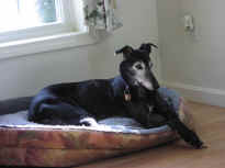
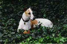
"Charming" 3/26/92-11/10/04 "Roxanne"
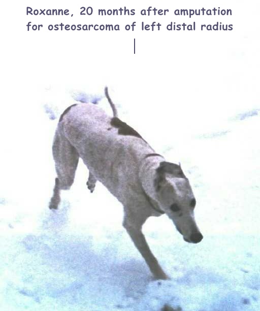
6/5/93-2/11/05
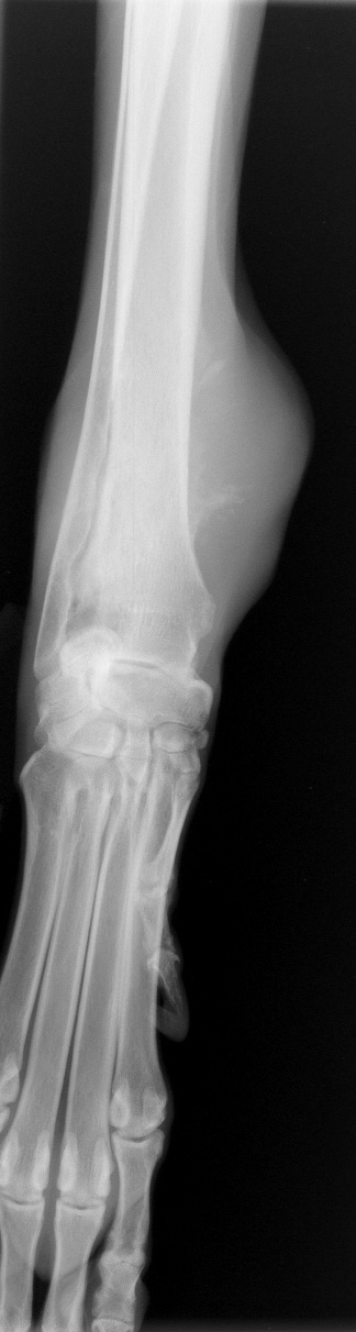
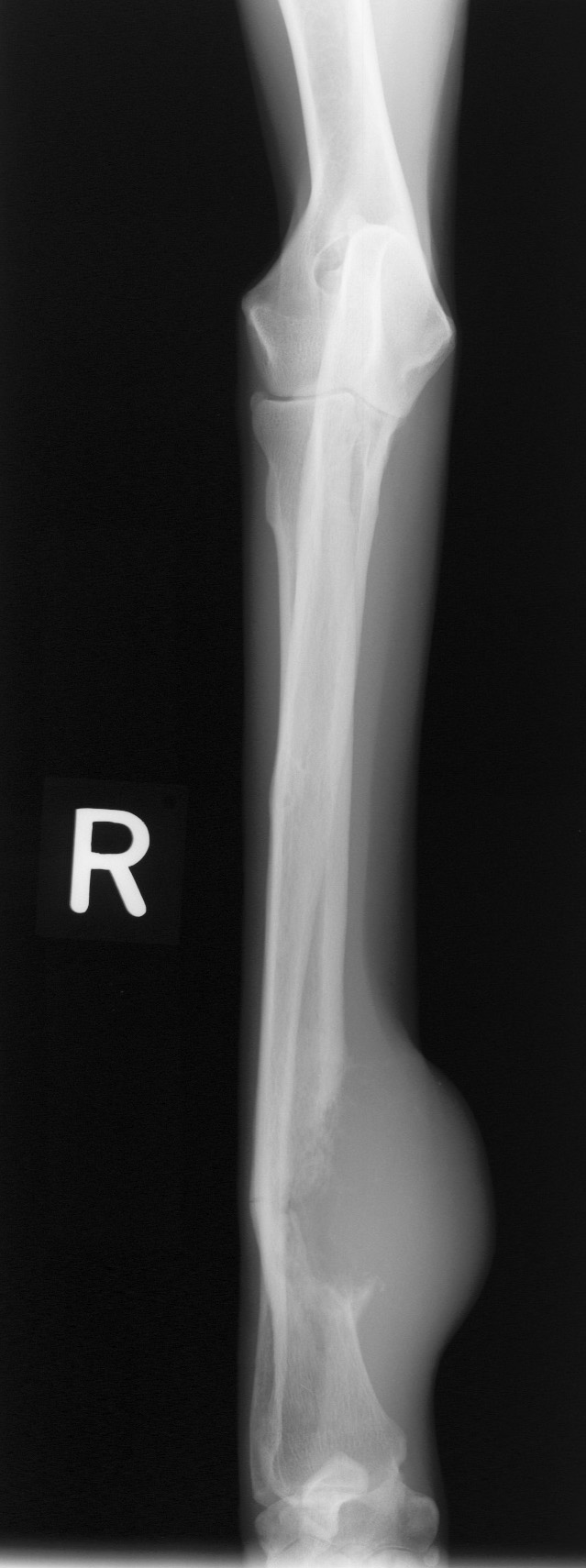
Osteosarcoma of Humerus Osteosarcoma of
Distal Radius
C. GUILLERMO COUTO,
DVM, Dip. ACVIM
Department of Veterinary
Clinical Sciences
College of Veterinary Medicine,
The Ohio State University, Columbus, OH 43210
Primary bone tumors (neoplasms) are common in dogs. Most primary
bone tumors in dogs are malignant, in that they usually cause death
as a result of local infiltration (e.g., pathologic fractures or
extreme pain leading to euthanasia) or dissemination (e.g.,
pulmonary metastases in osteosarcoma-OSA). Neoplasms that
metastasize (spread) to the bone are extremely rare in dogs; some
malignant tumors that occasionally metastasize to bones are
transitional cell carcinoma of the bladder, osteosarcoma in other
bone/s, hemangiosarcoma, mammary adenocarcinoma, and prostatic
adenocarcinoma.
Osteosarcomas are the
most common primary bone tumor in dogs, and the most common tumor in
Greyhounds in the United Kingdom, where it accounted for 50% of all
tumors, and for 22% of the deaths in the breed (www.gurk.demon.co.uk
/ghsurvey). Cancer in general (44%), and OSA in particular (22%)
were the leading cause of death in the breed. At the University of
Florida, 10% of all dogs with OSA were Greyhounds, and the risk of
developing OSA was higher for Greyhounds than for any other breeds.
OSAs
can affect either the appendicular (e.g.; legs) or axial (e.g.;
spine, skull) skeletons, and occur primarily in large (and
giant)–breed, middle age–to-older dogs. Preferential locations for
OSA include the distal radius, proximal humerus, and distal femur,
although they can occur in any bone or location; they are more
common in dogs over the age of 6 or 7.
Their biologic behavior is characterized by aggressive local
infiltration of the surrounding tissues and rapid dissemination
through the bloodstream (usually to the lungs). Although when first
discovered, most dogs with OSA have “clean” thoracic radiographs
(chest X-rays), there are usually tumor cells present in the lungs,
but the masses are too small to be seen in routine radiographs.
Clinical Signs (“Symptoms”)
OSAs
occur predominantly in the ends of the distal radius (wrist), distal
femur (knee), and proximal humerus (shoulder)(“TOWARDS
THE KNEE AND AWAY FROM THE ELBOW"), although other areas can
also be affected. In contrast with other breeds, where dogs with OSA
typically develop bone swelling and/or limping, Greyhounds
frequently develop a spontaneous pathological fracture (i.e.; the
bone breaks or fractures without any trauma).
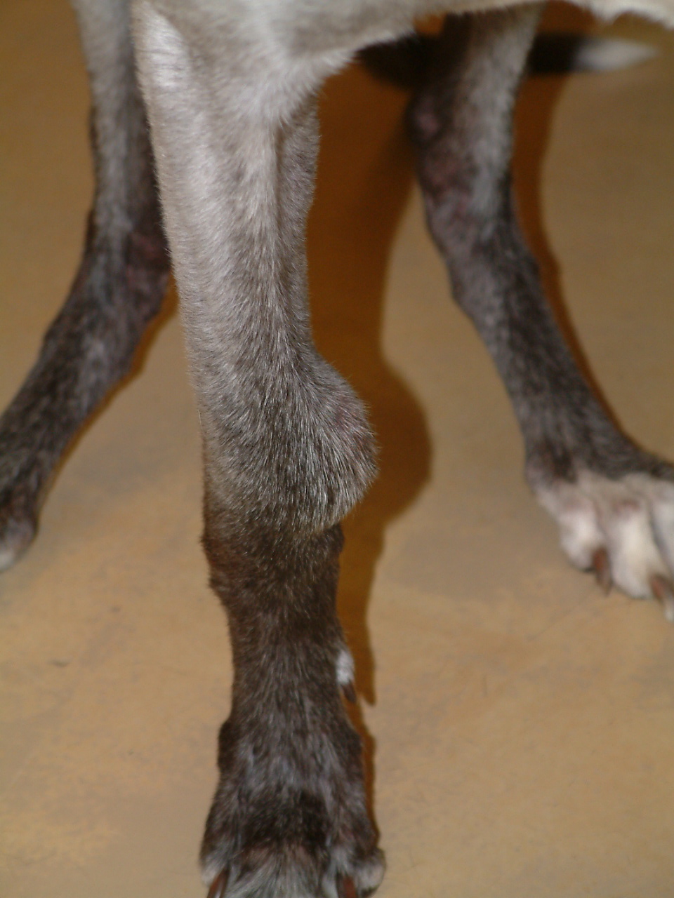
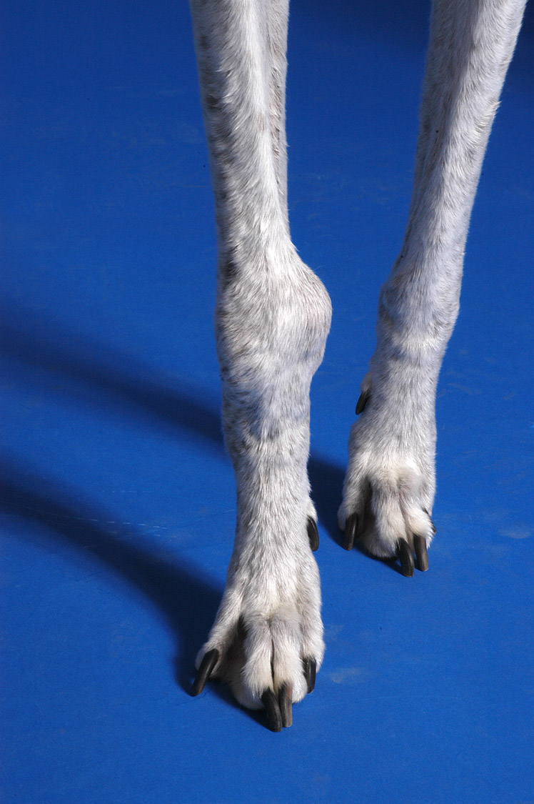
Osteosarcoma of distal radius in front leg
Diagnosis
 io
io
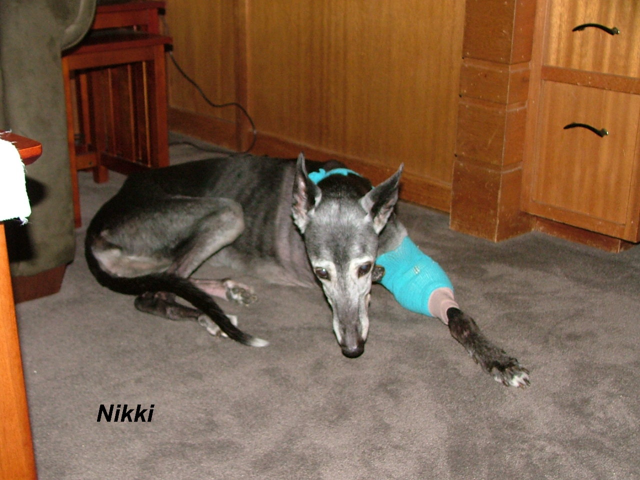
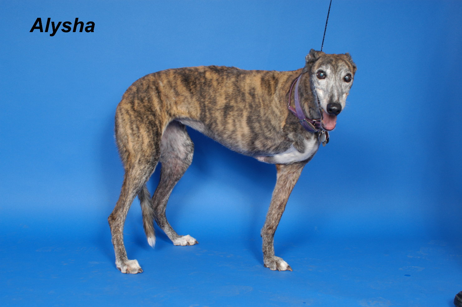
Destructive and proliferative bone changes in
dogs with OSA
(see
above photos for corresponding presentation
at time of examination)
If
the cytology is not diagnostic, and you are still debating whether
to go ahead with the limb amputation, a core biopsy of the affected
area should be obtained. For this, your veterinarian will use a
large bore needle with your hound under general anesthesia. The
diagnostic yield of this procedure is quite high (approximately 70%
to 75%), but because Greyhounds have very thin bones, the bone may
fracture (break) around the area of the biopsy.
As
long as you are aware of the biologic behavior of the tumor and the
clinical and radiographic features of the lesion are compatible with
OSA, the limb can be amputated in the absence of a biopsy. The
amputated leg (or representative samples) should always be submitted
for histopathologic studies.
Treatment and Prognosis
The
treatment of choice for dogs with OSA is amputation of the affected
limb, with adjuvant chemotherapy (another “bad word”-see below). The
median (average) survival time of dogs with appendicular OSA treated
with amputation alone is approximately 4 months, whereas in dogs
treated with amputation and chemotherapy it is approximately 1 year.
An
issue your veterinarian should be familiar with is the fact that
amputation in Greyhounds frequently results in severe postoperative
bleeding around the surgical site, leading to subcutaneous blood
accumulation in the other limbs, ventral thorax, and ventral
abdomen; your veterinarian should have access to plasma or other
blood products before the surgery. Alternatively, you can ask your
vet to refer you to a specialist (board-certified surgeon or
oncologist).
A
novel surgical approach for dogs with distal radial (wrist) OSA
consists of sparing the affected limb. Instead of amputation, the
affected bone is resected and an allograft from a cadaver is used to
replace the neoplastic bone; novel biomaterials are also currently
being investigated for this purpose. The dogs are also treated with
intravenous chemotherapy and, in general, have almost normal limb
function. The main complication is the development of osteomyelitis
(infection) in the allograft; if that occurs, the limb frequently
needs to be amputated. Survival times in dogs treated with
limb-sparing procedures are comparable to those in those that
undergo amputation plus chemotherapy, with the added benefit to the
owners of having a four-legged pet.
The
chemotherapeutic agents typically used for Greyhounds with OSA are
cisplatin, carboplatin, or doxorubin. The treatment results are
almost identical for the 3 drugs; because cisplatin has to be given
as an IV infusion, most oncologists are no longer using it. So,
basically, we are considering one of 2 conventional treatments: 5
doses of doxorubicin (Adriamycin) at 2-week intervals, or 4 doses of
carboplatin (Paraplatin) at 3-week intervals. Doxorubicin is
relatively inexpensive (a little bit over $120/dose for the cost of
the drug), whereas carboplatin is one of the most expensive chemo
drugs (approximately $40/kg of body weight, or an average of
$1,000/dose). However, carboplatin causes almost no side effects,
whereas approximately 20% of dogs receiving doxorubicin have mild
side effects, such as poor appetite, diarrhea, etc; also, dogs with
some types of heart disease cannot receive doxorubicin. These side
effects are minimal when compared with those in people on chemo.
And, oh, by the way, dogs on chemo don’t lose their hair. For the
average dog on chemo, there is no difference in the quality of life
when compared to that before chemo.
If
amputation is not an option, local radiotherapy plus chemo may be of
some benefit. However, in our limited experience, most dogs are
eventually euthanized within 3 to 4 months of the initial diagnosis
because of the development of pathological fractures (i.e., after
radiotherapy the tumor is not as painful; therefore the dog regains
normal use of the limb and fractures the area), osteomyelitis, or
metastatic lesions.
Pain
control is essential in dogs where surgery is not an option; we have
used either NSAIDs (carprofen, deracoxib, meloxicam) at recommended
doses, or bisphosphonates such as alendronate (Fosamax). Drugs such
as tramadol (Ultram) are also beneficial.
© C.
Guillermo Couto, DVM
couto.1@osu.edu
http://www.vet.ohio-state.edu/1872.htm
This article may not be
reproduced or published
without express consent of the author
couto.1@osu.edu
GreytHealth thanks Dr. Couto for his generosity in contributing this article, which will be an invaluable resource for owners of Greyhounds (and indeed all canine breeds) faced with this devastating diagnosis. The information Dr. Couto has provided will hopefully assist our readers in better understanding the diagnosis, treatment options, and prognosis of osteosarcoma. If you are concerned that your Greyhound may be displaying some of the signs of osteosarcoma described by Dr. Couto, PLEASE SEEK VETERINARY CARE IMMEDIATELY!

©
Copyright 2015 GreytHealth.com. All rights reserved.
Website by
WagWorks